MedNews

  • Drug Industry News
  • FDA News & Alerts
  • Health & Medical News
You are here: Home / Archives for Health & Medical News

Cognitive and Physical Decline in Older Adults Associated with Severe Sepsis

October 26, 2010 By MedNews Leave a Comment

Older adults who survive hospitalization involving severe sepsis, a serious medical condition caused by an overwhelming immune response to severe infection, are at higher risk for cognitive impairment and physical limitations than older adults hospitalized for other reasons, researchers have found.

The research, conducted by the University of Michigan and supported primarily by the National Institute on Aging (NIA), part of the NIH, appears in the Oct. 27, 2010, issue of the Journal of the American Medical Association.

Theodore J. Iwashyna, M.D., Ph.D., and colleagues found that an older person’s risk of cognitive decline increased almost threefold following hospitalization for severe sepsis. They also found that severe sepsis was associated with greater risk for the development of at least one new limitation in performing daily activities following hospitalization.

“Sepsis is common in older people and has a high mortality rate,” said NIA Director Richard J. Hodes, M.D. “This study shows that surviving sepsis may bring substantial and under-recognized problems with major implications for patients, families and the health care system.”

In sepsis, immune system chemicals released into the blood to combat serious infection trigger widespread inflammation. This can lead to low blood pressure, heart weakness, and organ failure. Anyone can get sepsis, but infants, children, older people, and those with weakened immune systems are most vulnerable. People with sepsis often receive treatment in hospital intensive care units to combat the infection, support vital organs and prevent a drop in blood pressure.

“This study should help change the way we think about severe sepsis,” said Iwashyna. “We usually think of severe sepsis as a medical emergency and focus our efforts on making sure the patient survives. This study shows that survivors often have severe problems for years afterwards.”

Using data from the NIA-supported Health and Retirement Study (HRS), the researchers analyzed the cognitive and physical function of older people before and after hospitalization for severe sepsis. The HRS is a long-term study that collects information on the health, economic and social factors influencing the health and well-being of a nationally representative sample of Americans over age 50. Study data on participants 65 and older are linked to Medicare claims data to enable detailed analysis of medical conditions and health status.

The scientists analyzed Medicare claims data from 516 people who survived 623 hospitalizations for severe sepsis between 1998 and 2005. The average age of participants was 77 at the time of hospitalization. The researchers also examined the individuals’ HRS data on cognitive function, measured through standard tests. Physical limitations were measured by the need for assistance in six activities of daily living basic self-care tasks (walking, dressing, bathing, eating, toileting and getting into and out of bed) and five instrumental activities of daily living (preparing a hot meal, shopping for groceries, making telephone calls, taking medicines and managing money), which are associated with the ability to live independently. For comparison, the researchers analyzed Medicare and HRS data on 4,517 survivors of 5,574 non-sepsis general hospitalizations during this time period.

Almost 60 percent of hospitalizations for severe sepsis were associated with worsened cognitive and/or physical function among survivors in the first survey following hospitalization. The risk of progression to moderate or severe cognitive impairment in sepsis survivors was 3.33 times higher than their risk before hospitalization. Severe sepsis was associated with the development of 1.57 new functional limitations among patients with no limitations before sepsis. In contrast, patients who did not develop sepsis and had no functional limitations before hospitalization developed an average of 0.48 new functional limitations. Non-sepsis hospital admissions were not associated with an increased risk for cognitive decline.

“This is one of more than a thousand research papers that have used Health and Retirement Study data,” said Richard Suzman, Ph.D., director of the NIA’s Division of Behavioral and Social Research, which supports the HRS. “The uniquely rich HRS dataset enabled the analysis of both cognitive and physical function in relation to hospitalization for a very specific medical condition. I look forward to the investigators refining their findings in the future.”

Source: NIH, October 26, 2010
Filed Under: Geriatrics, Mental Health Tagged With: dementia, geriatrics, seniors, sepsis

Study Finds Succimer Ineffective for Removing Mercury as Autism Treatment

October 22, 2010 By MedNews 2 Comments

Succimer, a drug used for treating lead poisoning, does not effectively remove mercury from the body, according to researchers. Some families have turned to succimer as an alternative therapy for treating autism.

“Succimer is effective for treating children with lead poisoning, but it does not work very well for mercury,” said Walter Rogan, M.D., head of the Pediatric Epidemiology Group at the National Institute of Environmental Health Sciences (NIEHS), part of NIH, and an author on the paper that appears online in the Journal of Pediatrics.

“Although it is not approved by the Food and Drug Administration to reduce mercury, succimer is reportedly being used for conditions like autism, in the belief that these conditions are caused, in part, by mercury poisoning,” Rogan stated. “Our new data offers little support for this practice.”

Although researchers found that succimer lowered blood concentrations of mercury after one week, continued therapy for five months only slowed the rate at which the children accumulated mercury. The safety of higher doses and longer courses of treatment has not been studied.

Most mercury exposure in the United States is from methylmercury, found in foods such as certain fish. Thimerosal, a preservative once more commonly used in vaccines, contains another form of mercury, called ethylmercury.

To conduct the study, the researchers used samples and data from an earlier clinical trial, led by NIEHS, called the Treatment of Lead-exposed Children (TLC) trial. In the TLC study, succimer lowered blood lead in 2-year-old children with moderate to high blood lead concentrations.

Using blood samples from 767 children who participated in the TLC trial, the researchers measured mercury concentration in the toddlers’ blood samples collected before treatment began, one week after beginning treatment with succimer or placebo, and then again after three month-long courses of treatment. Mercury concentrations were similar in all children before treatment. Concentrations eventually increased in both groups, but more slowly in the children given succimer. Succimer had produced a 42 percent difference in blood lead, but only an 18 percent difference in blood mercury.

“Although succimer may slow the increase in blood mercury concentrations, such small changes seem unlikely to produce any clinical benefit,” Rogan said. He and his colleagues had reported in an earlier paper that succimer has few adverse side effects, mostly rashes, and an unexplained increase in injuries in children given succimer rather than placebo.

The subjects of the study did not have unusually high blood mercury concentrations for African-American children and the study did not investigate where the mercury in the children came from.

“This research fills a gap in the scientific literature that could not be addressed any other way. We were fortunate to have samples already collected from toddlers who had been treated with succimer for lead poisoning allowing us to help answer this important question,” said Linda Birnbaum, Ph.D., director of NIEHS and the National Toxicology Program.

Reference: Cao Y, Chen A, Jones RL, Radcliffe J, Dietrich KN, Caldwell KL, et al. 2010. Efficacy of Succimer Chelation of Mercury at Background Exposures in Toddlers: A Randomized Trial. J Pediatr. Epub ahead of print. DOI:10.1016/j.jpeds.2010.08.036.

Source: National Institutes of Health (NIH), October 22, 2010

Filed Under: Autism, Pediatrics & Parenting Tagged With: autism, lead, mercury, sccimer

Body Cooling Treatment Studied for Pediatric Cardiac Arrest

October 19, 2010 By MedNews Leave a Comment

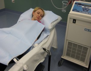
Blanketrol mattress and blanket used for body cooling.

The National Heart, Lung, and Blood Institute (NHLBI), part of the National Institutes of Health, has launched the first large-scale, multicenter study to investigate the effectiveness of body cooling treatment in infants and children who have had cardiac arrest. The Therapeutic Hypothermia after Pediatric Cardiac Arrest (THAPCA) trials total more than $21 million over six years.

Therapeutic hypothermia, or body cooling, has been successfully used in adults after cardiac arrest and in newborn infants after birth asphyxia, or lack of oxygen, to improve survival and outcomes, but it has not been studied in infants or children who have had cardiac arrest.

“Children who have experienced cardiac arrest can suffer long-term neurological damage or death,” said NHLBI Acting Director Susan B. Shurin, M.D., a board-certified pediatrician. “There are abundant data demonstrating the benefits of hypothermia in adults with cardiac arrest, but very limited experience in children. This study begins to assess the effectiveness of therapeutic hypothermia in children, and should lead to evidence-based guidelines that will optimize both quality and rates of survival.”

During body cooling treatment, THAPCA participants lie on mattresses and are covered with blankets. Machines circulate water through the blankets and mattresses to control the participants’ body temperatures. Researchers do not yet know how body cooling will affect participants, since many factors can contribute to brain injury after cardiac arrest. However, they believe body cooling could provide several benefits, including less inflammation and cell death.

According to a 2008 review of pediatric cardiopulmonary resuscitation in the journal Pediatrics, about 16,000 children suffer cardiac arrest each year in the United States. Their hearts stop pumping effectively, and blood stops flowing to their brains and other vital organs. In many cases, the outcome is death or long-term disability.

Cardiac arrest in infants and children has many causes, such as strangulation, drowning, or trauma. It can also be a complication of many medical conditions.

“Our goal is to minimize brain injury in infants and children who experience cardiac arrest and ultimately improve survival rates,” said co-principal investigator J. Michael Dean, M.D., M.B.A., professor of pediatrics and chief of the Division of Pediatric Critical Care Medicine at the University of Utah School of Medicine, Salt Lake City.

The THAPCA centers enroll participants in one of two randomized, controlled clinical trials. One evaluates participants who suffered cardiac arrest outside the hospital, while the other evaluates participants who suffered cardiac arrest in the hospital. Within each trial, there are two active treatment groups: therapeutic hypothermia (cooling the patient to 89.6-93.2 Fahrenheit) and therapeutic normothermia (maintaining the patient at 96.8-99.5 Fahrenheit). Both trials are trying to reduce fever, which commonly occurs after cardiac arrest and can lead to more severe outcomes.

“These trials are addressing the question: What is the optimal temperature for an infant or child after cardiac arrest?” said co-principal investigator Frank W. Moler, M.D., M.S., a professor in the Department of Pediatrics and Communicable Diseases at the University of Michigan, Ann Arbor. He added that in previous studies exploring therapeutic hypothermia, the comparison or control groups did not receive therapeutic normothermia to prevent fever.
Woman lying on a Blanketrol mattress and blanket used for therapeutic hypothermia.
Blanketrol mattress and blanket used for therapeutic hypothermia.

Participants in the THAPCA trials must be older than 48 hours and younger than 18 years and must be enrolled in the study within six hours of suffering cardiac arrest. Once a parent or guardian provides consent, the participant is randomly assigned to one of the two treatment groups. The therapeutic hypothermia group in each trial receives the hypothermia treatment for two days and then normothermia treatment for three days, which ensures that the body temperature is kept within a normal temperature range. The patients in the therapeutic normothermia groups receive normothermia treatment for all five days.

After the five-day period, the clinical care team will continue to provide study participants with optimal medical care. Participants will undergo neurological and behavioral testing a year after the cardiac arrest.

The THAPCA trials involve 34 clinical centers in the United States and Canada. The C.S. Mott Children’s Hospital at the University of Michigan serves as the lead clinical center, while the data coordinating center is based at the University of Utah School of Medicine.

Source: NIH, October 19, 2010

Filed Under: Heart Attack, Pediatrics & Parenting Tagged With: body cooling, heart attack, therapeutic hypothermia

Scientists Discover How Dengue Virus Infects Cells

October 12, 2010 By MedNews Leave a Comment

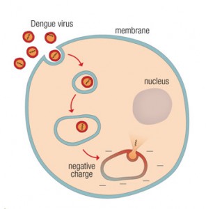
How does the dengue virus infect cells? To infect a cell, the dengue virus (counterclockwise, from upper left), binds to the cell membrane. The virus is then enveloped in the membrane, which coalesces around the virus, forming a pouch-like structure called an endosome. Deep inside the cell, the endosome membrane acquires a negative charge, which allows the virus to fuse with the endosomal membrane and release genetic material into the cell’s interior.

Researchers have discovered a key step in how the dengue virus infects a cell. The discovery one day may lead to new drugs to prevent or treat the infection.

Researchers from the National Institutes of Health discovered how the dengue virus releases itself from the protective membrane that shields it as it penetrates deep inside the cell. The discovery allows researchers to study the invasion process in the laboratory and provides a means to test potential treatments for the virus.

Dengue, which is transmitted by mosquitoes, infects up to 100 million people each year. People bitten by an infected mosquito first develop a fever, followed by other symptoms such as joint pain, rash and nausea. Without treatment, symptoms may become more severe. Patients with the severe form of the disease, dengue hemorrhagic fever, may develop difficulty breathing, bruising, bleeding from the nose or gums, and breakdown of the circulatory system. Each year, 22,000 people — most of them children — die from dengue, according to the World Health Organization.

To infect a cell, the virus binds to the cell membrane. The cell membrane engulfs the virus, enveloping it in a pouch-like structure known as an endosome. To begin the infection process, the virus delivers its hereditary material into the cytosol, the fluid interior of the cell, where it begins reproducing itself. To do so, however, it must first release itself from the endosome. The virus does this by fusing its membrane with the endosomal membrane. When the two membranes come together, they form a pore through which the virus’ genetic material is released.
Graphic shows how the virus is taken into the cell and releases its DNA in the presence of a negative charge.

To infect a cell, the dengue virus (counterclockwise, from upper left), binds to the cell membrane. The virus is then enveloped in the membrane, which coalesces around the virus, forming a pouch-like structure called an endosome. Deep inside the cell, the endosome membrane acquires a negative charge, which allows the virus to fuse with the endosomal membrane and release genetic material into the cell’s interior.

Scientists have used their understanding of HIV to develop drugs that block the fusion process and infection. To study the fusion stage of viral entry, researchers have typically observed viral fusion at the cell surface and fusion of a virus with an artificial membrane. Researchers working with dengue, however, were unable to get the virus to fuse under either of these conditions. Why dengue, unlike other viruses, would not readily fuse with these membranes had puzzled researchers for years.

The current study was undertaken by Leonid V. Chernomordik, Ph.D., of the Section on Membrane Biology at the NIH’s Eunice Kennedy Shriver National Institute of Child Health and Human Development and his colleagues, Elena Zaitseva, Sung-Tae Yang, Kamran Melikov, and Sergei Pourmal.

The researchers discovered that two conditions are necessary for dengue virus fusion—an acidic environment and the presence of a negatively charged membrane. They also discovered that these conditions are present at only certain points in the endosome’s journey within the cell.

“We spent several years trying to understand how the dengue virus fuses with its target membrane,” said Dr. Chernomordik. “The findings will now enable us to test new ways to disrupt the fusion process and prevent infection.”

Their research was published online in PLoS Pathogens.

To conduct their study, the researchers tagged dengue virus and cell membranes with molecules that would glow when the virus and membranes fused. They also exposed samples of the virus to an artificial membrane under various conditions, to identify factors that would allow fusion to take place.

The researchers first confirmed that a protein controlling fusion is active only under acidic conditions. However, conditions in the endosome are always acidic, and this alone was not enough to guarantee fusion, they found.

In additional experiments using artificial and cell membranes along with fluorescent markers, they discovered that fusion occurred only when the membranes were negatively charged. When the endosome begins its journey, the endosomal membrane has a neutral charge. The negative charge is present only after the endosome has been taken deep within the cell.

“The confluence of acidity and a negative charge deep in the cell’s interior ensures that the virus is safe within the endosome early in its journey, when it is most vulnerable, but can release its genome when it reaches its destination,” Dr. Chernomordik said.

Dr. Chernomordik and his colleagues plan to test various compounds to learn whether they can prevent the virus from fusing with cell and artificial membranes, in order to identify potential new treatments for dengue infection.

Source: NIH, October 12, 2010

Filed Under: Infectious Diseases Tagged With: dengue, NIH, research

Embryonic Stem Cell Research Testimony – Senate Subcommittee on Labor – HHS – Education Appropriations

September 27, 2010 By MedNews Leave a Comment

DEPARTMENT OF HEALTH AND HUMAN SERVICES
NATIONAL INSTITUTES OF HEALTH
The Promise of Human Embryonic Stem Cell Research
Witness appearing before the
Senate Subcommittee on Labor – HHS – Education Appropriations
Francis S. Collins, M.D., Ph.D.
Director, National Institutes of Health
September 16, 2010

Good morning, Chairman Harkin and distinguished Members of the Subcommittee. It is an honor to appear before you today to discuss human embryonic stem cell research. First, I’d like to thank this Subcommittee for its steadfast support of the National Institutes of Health’s (NIH) mission: discovering fundamental knowledge about living systems and then applying that knowledge to fight illness, reduce disability, and extend healthy life. NIH is grateful for the confidence that Congress – and this Subcommittee in particular – has shown in our ability to achieve this mission, as evidenced by our current $31 billion budget, and the $10.4 billion provided to NIH through the American Recovery and Reinvestment Act. Your support makes our mission possible, and we are very grateful.

Nowhere has this support been more evident than in this Subcommittee’s leadership in advancing human embryonic stem cell research. From your first hearing in December 1998, this Subcommittee has provided a forum for discussing the great promise this research holds. With your steadfast support, NIH has invested more than $500 million in human embryonic stem cell research; one of the most promising research avenues of recent times.

The preliminary injunction issued on August 23 by U.S. District Court Judge Royce Lamberth in the Sherley v. Sebelius case, now stayed pending further order from the Court of Appeals, has created uncertainty in the field of human embryonic stem cell research. Many researchers across the country have considered modifying their research plans to turn away from an area of research that, while promising, is now fraught with uncertainty. Some of our nation’s best researchers, who have written grant applications proposing innovative new ideas, are now asking, “Should I even bother to submit my
proposal to NIH?” Likewise, young scientists excited about careers in stem cell research are concerned about going into this field, given the legal uncertainty.

But the real reason for distress about the current legal uncertainty is that patients may have to put hope on hold. While we continue through the legal process, I hope that we can keep the patients and their families in our thoughts. They are at the heart of the NIH mission, and they are the ones who stand to benefit the most, or lose the most, by the stem cell policies we are discussing today.

I am not a lawyer, and I speak to you today as a doctor and a scientist. In that capacity, I want to outline for you the promise of human embryonic stem cell research — research that could be hobbled permanently unless stable federal funding can be assured over the long term.

I want to begin with a brief overview of the remarkable properties of human embryonic stem cells and then describe how research using these cells will:

  • provide key insights into the molecular pathways in development and disease;
  • allow for the development of tissue replacement or regenerative medicine; and
  • enable more targeted and efficient screening of new drug candidates.

Human Embryonic Stem Cells

Human embryonic stem cells possess several unique characteristics. First, these cells are pluripotent, which means that they have the potential to become nearly every one of the different types of cells in the human body. Second, these cells are self-renewing, which means that they are able to multiply in essentially limitless numbers in the lab over many years and to be shared with many researchers around the world.

To be sure, scientists are also interested in other types of stem cells. Adult stem cells are found in various organs and tissues throughout the body. These cells, also sometimes referred to as multipotent or somatic stem cells, can develop into a limited number of specific cell types, depending upon the organ or tissue from which they are derived. However, adult stem cells are less than ideal for many types of research and therapy because they do not divide indefinitely in culture, and they produce only a limited number of cells and cell types.

In considering the relative benefits of adult and embryonic stem cell research, keep in mind that research on the most abundantly available source of adult stem cells, hematopoetic stem cells in bone marrow, began more than a half-century ago. In fact, Drs. E. Donnall Thomas and Joseph Murray were awarded the Nobel Prize in Medicine in 1990, “for their discoveries concerning organ and cell transplantation in the treatment of human disease.” Indeed, this research has produced clinically-validated and widely-used treatments that reconstitute the immune system after leukemia, lymphoma, and various blood or autoimmune disorders have been treated with chemotherapy.

NIH is strongly committed to research using adult stem cells because there may be other clinical applications for which they prove useful. NIH has invested many hundreds of millions of dollars over the years in adult stem cell research. Indeed, annually we are spending almost three times as much on adult stem cell research as on human embryonic stem cell research.

A new and third category of stem cells are induced pluripotent stem (iPS) cells, which were created as a direct result of the knowledge gained from studying human embryonic stem cells.

This type of stem cell was first produced in 2007, when scientists discovered that it is possible to instruct adult skin cells to return to a very early developmental stage. They accomplished this by using viruses to insert molecular instructions into the DNA of skin cells – instructions that acted to turn back the cells’ developmental clock. These new cells possess many properties of human embryonic stem cells: they continue to divide indefinitely and are pluripotent, with the potential to give rise to all the cells of the human body.

While induced pluripotent stem cells are of great interest to scientists, and have the added potential clinical benefit of avoiding transplant rejection since they can be derived directly from the patient, they are not well understood yet. A growing body of research, including a publication just two months ago from Dr. George Daley, who is here today, and his collaborators suggests that there are subtle but potentially important differences between iPS cells and human embryonic stem cells. On close examination with powerful molecular fingerprinting, it seems that iPS cells retain some memory of the tissue from which they were derived. Whether this will matter for clinical applications is not clear, but virtually all investigators working in the field agree that additional comparisons between iPS cells and human embryonic stem cells are critically important. Human embryonic stem cells remain the gold standard for pluripotency: to prohibit work on human embryonic stem cells will thus do severe collateral damage to the new and exciting research on iPS cells.

Molecular Pathways in Biological Development and Human Disease

While many researchers are focused on coaxing human embryonic stem cells to develop into a particular cell type, such as insulin-secreting cells or liver cells, understanding the basic biology of stem cells themselves will be extremely valuable to understanding human development. We have learned much about the genes required for pluripotency, but there is much more to understand. For example, what genes are expressed in human embryonic stem cells? How is that program altered as these cells move down pathways to become blood cells, muscle cells, or brain cells? How are these steps regulated? What happens if one of the genes doesn’t function properly? Our best window into human development is using human embryonic stem cells.

In addition to understanding normal human development more completely, human embryonic stem cells are providing key tools to help us study the origins of many devastating diseases that afflict babies and young children. Such research may even help to uncover targets for drug development. We now have a number of human embryonic stem cell lines that are known to carry mutations that cause specific diseases. For example, scientists are studying cell lines with a mutation in the FMR1 gene that causes Fragile X, a developmental disability. The FMR1 gene normally makes a protein that the brain needs to develop properly. However, the Fragile X mutation in the FMR1 gene causes the body to make only a little or none of the protein. Research using human embryonic stem cells with this mutation showed that although the FMR1 gene is expressed normally in Fragile X, it is turned off after the cells begin to differentiate. How this happens is something we can study using human embryonic stem cell lines. Dr. Daley also studies a number of human embryonic stem cell lines with various genetic mutations, and I am sure he can tell you more about his research.

One ongoing NIH grant focuses on Rett syndrome, a debilitating, developmental brain disorder generally afflicting young females and caused by mutations in a gene called MECP2. This research uses human embryonic stem cells to generate human brain cells with a deficiency in the MECP2 gene, and then studies the effects of this deficiency on the development and functions of these brain cells. Such research could improve our understanding of Rett syndrome, and facilitate the development of therapies for it.

Another research team has recently generated human embryonic stem cell lines containing mutations in the HTT gene that causes Huntington’s disease, a late-onset neurodegenerative disease. Huntington’s disease has been studied for a long time, but the normal function and pathogenesis of the protein coded for by the HTT gene is not fully understood.

Tissue Replacement or Regenerative Medicine

One of the more exciting and high-profile potential applications of human embryonic stem cell research is the possibility that such cells can be programmed to replace or regenerate tissues damaged by disease or injury. For example, we might one day be able to regenerate damaged heart muscle tissue in heart attack patients, develop insulin-producing pancreatic beta cells to replace those lost or damaged in people with Type 1 diabetes, or restore spinal cord neural connections in patients paralyzed by catastrophic spinal cord injury.

Part of the devastation that heart attack victims suffer is that, because of restricted blood flow and oxygen deprivation, their heart muscle cells die, leaving the heart much weaker and less able to pump blood throughout the body. Today we are studying the tantalizing possibility that human embryonic stem cells, or perhaps adult stem cells or iPS cells, might be programmed to replace damaged or destroyed heart muscle cells, known as myocardial cells. The prevalence of heart disease, along with the scarcity of hearts and heart tissues available for transplantation and the associated clinical and autoimmune problems of transplantation, make this line of research imperative.

Type 1 diabetes is a disease in which a specific type of cell, insulin-producing pancreatic beta cells, is damaged or destroyed by the patient’s own immune system. A major challenge is to understand the autoimmune response that kills these cells in children who then develop Type 1 diabetes, but human embryonic stem cells offer the hope that we might one day produce replacement cells that avoid the autoimmune challenges associated with today’s rudimentary transplantation therapies. To do that, we need to know more about how stem cells are genetically programmed, how they differentiate, and how they renew themselves; but as our understanding and ability to work with these cells expands, we are laying the foundation for an entirely new — and much more effective — way to address the devastation of Type 1 diabetes.

One of the most exciting — and most advanced — possible therapeutic applications of human embryonic stem cells is for patients who have been paralyzed by catastrophic spinal cord damage. Researchers at the University of California-Irvine and at the biotech company Geron Corp., as well as at other universities and companies around the country, are pursuing the possibility that human embryonic stem cells can be directed to generate spinal cord cells for transplantation.

This summer, Geron began Phase I safety trials of its technique for converting stem cells into a type of neuronal cell, known as oligodendrocytes, intended for injection into the patient’s spinal cord at the site where it has been severed by injury. The hope, which has been repeatedly demonstrated in animal tests, is that the newly-injected oligodendrocytes might repair the damaged insulation around the severed nerve cells of the spinal cord, and thereby enable those cells to once again send signals to the patient’s limbs and organs. We are not sure that this approach will work, and even if it does, it will take years of additional research and testing before we can develop a standardized therapy using these techniques. Still, the potential that this research holds is truly amazing.

For all of these efforts, there are many scientific challenges that must be addressed. We need to figure out how to get human embryonic stem cells to differentiate down specific pathways in a well-controlled process. We also need to make sure that the resulting cells behave in predictable ways. Because human embryonic stem cells are immortal and can proliferate endlessly — much like cancer cells — we need to be sure that they or their differentiated “daughter” cells do not produce tumors or otherwise harm patients. The field of regenerative medicine is young. It is unreasonable for us to think we will have cures within a set time period. It is also wrong to overpromise on the speed and scope of such research to patients and their families. But we must persevere and move this research forward in a strong and consistent manner. That is why the delay and uncertainty associated with the current legal situation is so disheartening for both researchers and patients. As I said at the time the injunction was issued, this unexpected development is like pouring sand into the engine of discovery.

Targeted, Efficient Screening of New Drug Candidates

Recently, human embryonic stem cells have received increasing attention as a tool for drug screening. High throughput drug screening is done in a miniaturized format that allows researchers to test the effect of more than 100,000 chemicals on a gene, protein, cell, or organism of interest. It is a highly automated process that can test in one day what would otherwise take a researcher months or years. Because human embryonic stem cells can differentiate into specific human cell types in large quantities, they provide the foundation for high throughput screening of candidate drug compounds for a given disease. This means that we now have the capacity to identify efficiently drugs that work in a targeted cell type.

This is not a promise, it is reality. Human embryonic stem cells are currently being used to identify drug candidates that can slow or stop the progression of amyotrophic lateral sclerosis (ALS). Also called Lou Gehrig’s disease, ALS is an ultimately fatal disease characterized by the progressive loss of motor neurons, which provide the connection between the brain and the muscles of our body. The possibility that human embryonic stem cell research might one day enable us to identify a therapy for the disease that afflicts astrophysicist Stephen Hawking and claimed the life of Senator Jacob Javits, gives you some sense of the hope this new application might provide.

There are very few drugs available for ALS, and none that prolong the patient’s life for more than a few months. Dr. Lee Rubin, a researcher at Harvard’s Stem Cell Institute, and his colleagues have developed an elegant set of studies to screen for drugs that prevent motor neuron death. The scientists differentiated mouse embryonic stem cells into large numbers of motor neurons and exposed them to thousands of compounds to find the ones that improve the survival of these vital cells. Dr. Rubin and his team identified a handful of promising compounds that they then tested in motor neurons derived from human embryonic stem cells. The most promising of these can now be moved further along the pipeline from drug discovery to clinical trials.

Drugs fail for many reasons: lack of efficacy in humans is responsible for 30% of drug failures, and unpredicted toxicity is responsible for more than 20% of failures. The traditional methods of using animal or abnormal human cell lines for safety and efficacy testing provide a poor model of how a human will respond to a particular drug. Human embryonic stem cells can generate the appropriate cell type and even disease cell lines for efficacy testing early on, as in the case of the ALS study. They are also being used to understand the toxicity of promising compounds in the early stages of drug development. For example, liver toxicity is a common cause of drug failure. Human embryonic stem cells can be differentiated into human liver cells, or hepatocytes, which are then exposed to novel drugs to identify any obvious liver toxicity and provide early insight on how a drug will be metabolized by the liver. In this manner, human embryonic stem cells provide drug developers and researchers a model of how actual human livers will respond to a drug. Our hope is this will reduce the number of drugs that fail in human clinical trials because of low efficacy or unacceptable toxicity.

The NIH Stem Cell Guidelines

President George W. Bush first funded research on human embryonic stem cells – but that decision only allowed the use of cell lines that had been derived before August 9, 2001. Ultimately, that only amounted to 21 cell lines, and as science moved forward it was clear that this somewhat arbitrary time stamp was significantly inhibiting the field. On March 9, 2009, President Barack Obama issued Executive Order 13505, Removing Barriers to Responsible Scientific Research Involving Human Stem Cells. The Executive Order states that the Secretary of Health and Human Services, through the Director of NIH, may support and conduct responsible, scientifically worthy human stem cell research, including human embryonic stem cell research. This Executive Order prompted a rapid expansion of scientific effort and progress.

The President asked NIH to review existing human stem cell research guidelines and issue new guidelines, consistent with the President’s Executive Order, within 120 days. NIH immediately began a comprehensive review that resulted in draft guidelines that were published in the Federal Register for public comment on April 23, 2009. After careful analysis of more than 49,000 comments from scientific, patient advocacy, medical, and religious organizations, as well as private citizens and members of Congress, NIH published final guidelines, effective July 7, 2009. The guidelines provide a framework for funding scientifically worthy research using responsibly derived human embryonic stem cells. The guidelines restrict federal funding to cell lines derived from embryos that: were created for reproductive purposes and were no longer needed for that purpose; were donated for research by individuals who sought reproductive treatment;

and for which the donors gave voluntary written consent. Since the President issued his Executive Order and NIH implemented its guidelines, 75 human embryonic stem cell lines have been approved for use in NIH-funded research. All were reviewed rigorously and found to meet the high ethical standards laid out in the NIH guidelines. The review process is so rigorous that 48 stem cell lines have not been approved for use in federally funded research. Prior to the court’s order, in FY 2010, NIH funded 199 grants for research on human embryonic stem cells totaling $137 million. These grants support a broad range of research including studies to improve stem cell technologies, studies to compare different types of stem cells, and studies to develop cell types for use in treating debilitating diseases and disorders such as diabetes, liver failure, and neurodegenerative diseases.

If the government is not successful in defending the guidelines in this litigation, and NIH will have to withdraw future NIH support for all grants involving human embryonic stem cell research, drastic scientific consequences will occur. Since funding for these projects would be discontinued mid-stream, all the funds that have been put in accounts or already drawn down — $270 million over the two- to five-year life of these grants, including what has been provided FY 2010 – would have been wasted. The research momentum that this Subcommittee worked so hard to achieve would be lost.

Young scientists may opt out of this field due to the chaos of stopping, then starting and now stopping again. More senior investigators may look to other countries, such as Singapore, China, and the United Kingdom to pursue their work. The greatest loss, however, will be for the millions of Americans with conditions currently under study with human embryonic stem cells. Such people include those suffering from heart disease, diabetes, liver disease, and vision problems, along with those afflicted by spinal cord injuries and neurodegenerative conditions like ALS and Alzheimer’s disease. The many messages I have received from patients since the issuance of the preliminary injunction reflect these deep concerns. Here is part of just one such message:

“I am a mother of two adult sons with Type I diabetes (since age 7), and a person with young onset Parkinson’s disease. I have watched as my oldest son moved from taking the old beef/pork insulin to taking genetically engineered insulin, and have held my breath with hope that my sons would benefit from the early stem cell research.

I watched as American scientists and science fell farther behind on the global scene during the past decade. In 2009 I had such hope that once again our medical schools and universities would begin to attract the best and brightest young minds to work in this exciting and promising area of research.

This week’s news was devastating to me. I had no idea how strongly I would be affected by it. Your message of support for the research once again gives me hope. Hope that there will be change. Hope that we will see effective treatments in our lifetimes for these devastating diseases.”

Thank you, Mr. Chairman, for your strong support of stem cell research. I would be
happy to answer any questions.

Francis S. Collins, M.D., Ph.D.

Francis S. Collins, M.D., Ph.D., was officially sworn in on Monday, August 17, 2009 as the 16th director of the National Institutes of Health (NIH). Dr. Collins was nominated by President Barack Obama on July 8, and was unanimously confirmed by the U.S. Senate on August 7.

Dr. Collins, a physician-geneticist noted for his landmark discoveries of disease genes and his leadership of the Human Genome Project, served as director of the National Human Genome Research Institute (NHGRI) at the NIH from 1993-2008. With Dr. Collins at the helm, the Human Genome Project consistently met projected milestones ahead of schedule and under budget. This remarkable international project culminated in April 2003 with the completion of a finished sequence of the human DNA instruction book.

In addition to his achievements as the NHGRI director, Dr. Collins’ own research laboratory has discovered a number of important genes, including those responsible for cystic fibrosis, neurofibromatosis, Huntington’s disease, a familial endocrine cancer syndrome, and most recently, genes for type 2 diabetes and the gene that causes Hutchinson-Gilford progeria syndrome.

Dr. Collins has a longstanding interest in the interface between science and faith, and has written about this in The Language of God: A Scientist Presents Evidence for Belief (Free Press, 2006), which spent many weeks on The New York Times bestseller list. He is the author of a new book on personalized medicine, The Language of Life: DNA and the Revolution in Personalized Medicine (HarperCollins, 2010).

Dr. Collins received a B.S. in chemistry from the University of Virginia, a Ph.D. in physical chemistry from Yale University, and an M.D. with honors from the University of North Carolina at Chapel Hill. Prior to coming to the NIH in 1993, he spent nine years on the faculty of the University of Michigan, where he was a Howard Hughes Medical Institute investigator. He is an elected member of the Institute of Medicine and the National Academy of Sciences. He is the recipient of the Presidential Medal of Freedom (2007) and the National Medal of Science (2009). On April 22, 2010, Dr. Collins was a co-recipient of the Albany Medical Center Prize in Medicine and Biomedical Research.

Filed Under: Health & Medical News, Stem Cells Tagged With: HHS, stem cells

Study Models H1N1 Flu Spread

September 21, 2010 By MedNews Leave a Comment

As the United States prepares for the upcoming flu season, a group of researchers continues to model how H1N1 may spread.

The work is part of an effort  called the Models of Infectious Disease Agent Study (MIDAS), to develop computational models for conducting virtual experiments of how emerging pathogens could spread with and without interventions. The study, supported by the National Institutes of Health, involves more than 50 scientists with expertise in epidemiology, infectious diseases, computational biology, statistics, social sciences, physics, computer sciences and informatics.

As soon as the first cases of H1N1 infections were reported in April 2009, MIDAS researchers began gathering data on viral spread and affected populations. This information enabled them to model the potential outcomes of different interventions, including vaccination, treatment with antiviral medications and school closures. The work built upon earlier models the MIDAS scientists developed in response to concerns about a different potentially pandemic influenza strain H5N1 or avian flu.

“Computational modeling can be a powerful tool for understanding how a disease outbreak is unfolding and predicting the implications of specific public health measures,” said Jeremy M. Berg, Ph.D., director of the National Institute of General Medical Sciences, which supports MIDAS. “During the H1N1 pandemic, MIDAS scientists applied their models to see what they could do to help in a real situation.”

Because the H1N1 flu strain is still circulating, a MIDAS group based at the University of Washington in Seattle is now studying the impact the virus could have this fall and winter. Its model, which represents the world population, includes information about immunity — how many people are protected by vaccination or prior infection—and the other circulating flu strains. Using the model, the scientists may be able to predict how H1N1 evolves and the possible role of the H3N2 strain, which historically has been the dominant seasonal flu virus. The results also may help forecast the potential effectiveness of the new flu vaccine that includes both the H1N1 and H3N2 viral strains.

Estimating Severity

To predict the likely severity of H1N1 in the fall and winter months following the initial outbreaks, the MIDAS group led by Marc Lipsitch, D.Phil., of the Harvard School of Public Health in Boston analyzed patient care data from Milwaukee and New York City. The researchers estimated that about 1 in 70 symptomatic people were admitted to the hospital, 1 in 400 required intensive care and 1 in 2,000 died. They predicted H1N1 to be no more and possibly even less severe than the typical seasonal flu strain. The work, which factored in local differences in flu detection and reporting, also showed that it’s possible to make predictions about severity using data from the early stages of an outbreak.

Vaccinating Children

Ira Longini, Ph.D., at the University of Washington and his MIDAS colleagues developed a simulation model to evaluate the effectiveness of different strategies to vaccinate school-aged children, who are known to play a key role in transmitting the flu virus. They modeled a range of scenarios that varied the type of vaccine, the percentage of children vaccinated and the infectiousness of the virus. For each situation, the modeling results indicated that vaccinating this age group substantially reduced overall disease spread and prevented up to 100 million additional cases in the general population. These effects, however, were less strong when the virus was more contagious or when fewer children were vaccinated. Based on these results, Longini’s group concluded that vaccine distribution strategies should depend on a number of factors, including vaccine availability and viral transmission rates.

Cost-Benefit of Employee Vaccination Programs

In one of the first analyses of the economic value of work-sponsored seasonal and pandemic flu vaccine programs, the MIDAS group led by Donald Burke, M.D., at the University of Pittsburgh developed a model that estimated the employer cost to be less than $35 per vaccinated employee with a potential savings of $15 to $1,494 per employee, depending on the infectiousness of the virus.

Interventions and Local Demographics

To determine if a vaccination strategy would likely have the same effect in different locations, a team led by MIDAS investigator Stephen Eubank, Ph.D., of the Virginia Bioinformatics Institute at Virginia Tech in Blacksburg developed models representing the demographics of Miami, Seattle and each county in Washington. The models indicated that while vaccinating school-aged children was the best strategy in each place, the optimal timing and overall effectiveness of the approach varied due to specific characteristics of the local population, such as age, income, household size and social network patterns. These differences, Eubank concluded, suggest that vaccination and probably other intervention strategies should take local demographics into account.

Antiviral Medications

Lipsitch’s collaborators Joseph Wu, Ph.D., and Steven Riley, D.Phil., at the University of Hong Kong used mathematical modeling to predict the likelihood that the H1N1 strain would develop resistance to the widespread use of antiviral medications taken to lessen flu symptoms. Their work showed that giving a secondary antiviral flu drug either prior to or in combination with a primary antiviral could mitigate the emergence of resistant strains in addition to slowing the spread of infection. The results, the researchers concluded, point to the value of stockpiling more than one type of antiviral drug.

School Closures

A public health measure under consideration was closing schools, which previous MIDAS pandemic flu models identified as a potentially effective intervention. According to Burke’s model of Allegheny County, Penn., closing individual schools after they identified cases may work as well as closing entire school systems. When strictly maintained for at least 8 weeks, both types of school closure could delay the epidemic peak by up to 1 week, allowing additional time to develop and implement other interventions. However, the model also indicated that school closures lasting less than 2 weeks could actually facilitate flu spread by returning susceptible students to school in the middle of an outbreak.

“Models like the ones MIDAS has developed help us understand not only trends in disease spread, but also how different factors can influence those trends,” said Irene A. Eckstrand, Ph.D., who directs the MIDAS program. “MIDAS research is leading to new tools and approaches that can aid in making public health decisions at a range of levels, from local to national.”

Source: National Institutes of Health (NIH), September 21, 2010

Filed Under: Infectious Diseases Tagged With: flu, h1n1, swine flu

20,000+ Annual Foodborne Illnesses Reported, Says CDC

August 12, 2010 By MedNews Leave a Comment

A total of 1,097 foodborne disease outbreaks were reported in 2007 to the Centers for Disease Control and Prevention, according to a CDC analysis. State investigators reported 21,244 illnesses and 18 deaths as a result of these outbreaks. The report also provides the most recent data on how many illnesses were linked to specific types of foods.

“Knowing more about what types of foods and foodborne agents have caused outbreaks can help guide public health and the food industry in developing measures to effectively control and prevent infections and help people stay healthy,” said Chris Braden, acting director of the CDC’s Division of Foodborne, Waterborne and Environmental Diseases.

Despite health officials’ efforts, the cause of an outbreak—either the food or the foodborne agent responsible—often cannot be determined or confirmed. This most commonly is the case when the outbreak is small. Of 1,097 reported outbreaks in 2007, 497 (or 45 percent) confirmed that one foodborne agent was responsible and in an additional 12 outbreaks more than one foodborne agent was responsible. Thus, in more than half of the outbreaks, a foodborne agent was not identified. Norovirus was the most frequently confirmed foodborne agent (39 percent), followed by Salmonella (27 percent).

Foodborne disease outbreaks due to norovirus occur most often when infected food handlers do not wash their hands well after using the toilet; outbreaks due to salmonella occur most often when foods are contaminated with animal feces. Contaminated foods are often of animal origin, such as beef, poultry, milk, or eggs. But any food, including vegetables, may become contaminated. Thorough cooking kills Salmonella.

The report states that in the 235 outbreaks where one food commodity was identified, the largest number of illnesses listed poultry (691 illnesses), beef (667 illnesses), and leafy vegetables (590 illnesses) as the cause. The CDC tracks 17 food commodity categories.

To prevent foodborne illnesses, CDC recommends that consumers and food handlers appropriately clean, separate, cook and chill foods.

Source: CDC, August 12, 2010

Filed Under: Food Tagged With: food illness, food safety, statistics

World Health Organization Declares End to H1N1 Influenza Pandemic

August 10, 2010 By MedNews Leave a Comment

The World Health Organization (WHO) International Health Regulations (IHR) Emergency Committee and the WHO Director-General, Dr. Margaret Chan, today declared an end to the 2009 H1N1 influenza pandemic. This declaration was based on strong indications that influenza, worldwide, is transitioning toward seasonal patterns of transmission.

In the majority of countries, out-of-season 2009 H1N1 outbreaks are no longer being observed, and the intensity of 2009 H1N1 influenza virus transmission is lower than that reported during 2009 and early 2010. Members of the Emergency Committee further noted that the 2009 H1N1 viruses will likely continue to circulate for some years to come, taking on the behavior of a seasonal influenza virus.

This does not mean that the H1N1 virus has disappeared. Rather, it means current influenza outbreaks including those primarily caused by the 2009 H1N1 virus, show an intensity similar to that seen during seasonal epidemics. Pandemics, like the viruses that cause them, are unpredictable. WHO noted that continued vigilance is extremely important, and it is likely that the virus will continue to cause serious disease in younger age groups and pregnant women, at least in the immediate post-pandemic period.

The WHO Director-General ended the Public Health Emergency of International Concern in accordance with the International Health Regulations (2005).

Implications for United States
This is a formal WHO declaration regarding the end of the pandemic at the global level. The U.S. Public Health Emergency determination for 2009 H1N1 Influenza expired on June 23, 2010.

The only impact on the United States resulting from the WHO declaration will be a cessation in weekly reporting under the International Health Regulations (IHR) to the Pan American Health Organization and the World Health Organization. CDC has reported weekly to IHR since early in the pandemic.

There are no changes for the United States in terms of CDC’s recommendations for the upcoming influenza season and the United States is already proceeding with the understanding that the 2009 H1N1 virus is now part of seasonal influenza virus circulation.

Protecting Yourself and Others from Influenza
CDC recommends a three-step approach to fighting flu: vaccination, everyday preventive actions and the correct use of antiviral drugs if your doctor recommends them. The first and most important step in protecting against the flu is to get a flu vaccine each season.

The U.S. 2010-2011 influenza vaccine will protect against an H3N2 virus, an influenza B virus, and the 2009 H1N1 influenza virus that caused the first global pandemic in more than 40 years and resulted in substantial illness, hospitalizations and deaths. In the United States, the CDC’s Advisory Committee on Immunization Practices recently recommended that everyone 6 months of age and older be vaccinated against influenza each season. Pregnant women, young children, and anyone with underlying health conditions like asthma, diabetes and neuromuscular diseases are at especially high risk for influenza-related complications and, therefore, should be vaccinated as soon as vaccine becomes available. Vaccine manufacturers are predicting an ample supply of influenza vaccine for the upcoming 2010-2011 U.S. influenza season.

Source: CDC, August 10, 2010

Filed Under: Infectious Diseases Tagged With: flu, h1n1, swine flu, world health organization

U.S. Adult Obesity Rates on the Rise

August 3, 2010 By MedNews Leave a Comment

The number of states with an obesity prevalence of 30 percent or more has tripled in two years to nine states in 2009, according to a CDC Vital Signs report. In 2000, no state had an obesity prevalence of 30 percent or more. The report, “State-Specific Obesity Prevalence Among Adults – United States, 2009,” also finds no state met the nation’s Healthy People 2010 goal to lower obesity prevalence to 15 percent.

The data show a 1.1 percentage point increase—an additional 2.4 million people—in the self-reported prevalence of obesity between 2007 and 2009 among adults aged 18 and over. The report also notes the medical costs associated with obesity are high. In 2008 dollars, medical costs associated with obesity were estimated at $147 billion. People who are obese had medical costs that were $1,429 higher than those of normal weight, the report said.

“Obesity continues to be a major public health problem,” said CDC Director Thomas Frieden, M.D., M.P.H. “We need intensive, comprehensive and ongoing efforts to address obesity. If we don’t more people will get sick and die from obesity-related conditions such as heart disease, stroke, type 2 diabetes and certain types of cancer, some of the leading causes of death.”

The August Vital Signs report is based on new data from the Behavioral Risk Factor Surveillance System (BRFSS). BRFSS contains state-level public health data and provides a way for states to monitor progress toward Healthy People goals. To assess obesity prevalence, approximately 400,000 phone survey respondents were asked to provide their height and weight, which was used to calculate their body mass index (BMI). An adult is considered obese if he or she has a BMI of 30 or above. For example, a 5-foot-4 woman who weighs 174 pounds or more, or a 5-foot-10 man who weighs 209 pounds or more has a BMI of 30, and so is considered obese.

The BRFSS obesity data are underestimates of true obesity prevalence. Research has found that both men and women often say they are taller than they actually are and women often say they weigh less than they do in telephone surveys. As a result, according to William Dietz, M.D., Ph.D., director of CDC’s Division of Nutrition, Physical Activity and Obesity, the overall BRFSS obesity prevalence estimate of 26.7 percent is 7.2 percentage points lower than the national 2007-2008 estimate of 33.9 percent (nearly 73 million people) from the National Health and Nutrition Examination Survey, for which individuals’ height and weight were measured rather than self-reported.

The BRFSS data highlight how obesity affects some populations more than others. The highest prevalence was found among non-Hispanic blacks overall, whose rate was 36.8 percent, and non-Hispanic black women, whose rate was 41.9 percent. The rate for Hispanics was 30.7 percent. The rate among all non-high school graduates was 32.9 percent. Obesity prevalence was also higher in some regions than others. The South had an obesity prevalence of 28.4 percent while the Midwest had a prevalence of 28.2 percent.

“Obesity is a complex problem that requires both personal and community action,” said Dr. Dietz. “People in all communities should be able to make healthy choices, but in order to make those choices there must be healthy choices to make. We need to change our communities into places where healthy eating and active living are the easiest path.”

Filed Under: Diet & Weight Tagged With: obesity, statistics, USA

90% of U.S. Adults Get Too Much Salt

June 24, 2010 By MedNews Leave a Comment

Less than 10 percent of U.S. adults limit their daily sodium intake to recommended levels, according to a new report, “Sodium Intake in Adults – United States, 2005-2006,” published today in Morbidity and Mortality Weekly Report. The report also finds that most sodium in the American diet comes from processed grains such as pizza and cookies, and meats, including poultry and luncheon meats.

According to the report, U.S. adults consume an average of 3,466 milligrams (mg) of sodium per day, more than twice the current recommended limit for most Americans. Grains provide 36.9 percent of this total, followed by dishes containing meat, poultry, and fish (27.9 percent). These two categories combined account for almost two-thirds of the daily sodium intake for Americans.

An estimated 77 percent of dietary sodium comes from processed and restaurant foods. Many of these foods, such as breads and cookies, may not even taste salty. “Sodium has become so pervasive in our food supply that it’s difficult for the vast majority of Americans to stay within recommended limits,” said Janelle Peralez Gunn, public health analyst with CDC’s Division for Heart Disease and Stroke Prevention and lead author of the report. “Public health professionals, together with food manufacturers, retailers and health care providers, must take action now to help support people’s efforts to reduce their sodium consumption.”

The 2005 Dietary Guidelines for Americans recommends that people consume less than 2,300 mg of sodium per day. Specific groups, including persons with high blood pressure, all middle-aged and older adults and all blacks, should limit intake to 1500 mg per day. These specific groups comprise nearly 70 percent of the U.S. adult population. This study found that only 9.6 percent of all participants met their applicable dietary recommendation, including 5.5 percent of the group limited to 1,500 mg per day and 18.8 percent of the 2,300 mg per day group.

The report examined data for 2005–2006 from the National Health and Nutrition Examination Survey (NHANES), an ongoing study that explores the health and nutritional status of adults and children in the United States. Researchers used information from 24-hour dietary recall and the USDA National Nutrient Database to estimate the daily sodium intake and sources of sodium intake for U.S. adults.

The findings add to a growing body of observational research studies on Americans’ excessive sodium consumption. Overconsumption of sodium can have negative health effects, including increasing average levels of blood pressure. One in three U.S. adults has high blood pressure, and an estimated 90 percent of U.S. adults will develop the disease in their lifetime. Blood pressure is a major risk factor for heart disease and stroke, the first and third leading causes of death among adults in the United States.

Source: CDC, June 24, 2010

Filed Under: Diet & Weight Tagged With: diet, salt, statistics, USA

« Previous Page
Next Page »

Search


Tag Cloud

alcohol allergies alzheimers antibiotics breast cancer cancer Centers for Disease Control (CDC) children cholesterol dementia depression Diabetes diet drug abuse exercise fda flu food genetics geriatrics h1n1 heart heart attack heart disease high blood pressure HIV/AIDS hormone therapy hypertension lungs menopause NIH obesity pain pediatrics pregnancy prostate cancer seniors sleep smoking statistics stroke sun teens USA women's health

Recent Comments

  • Mia Fritz on Health Benefits of Dark Chocolate
  • Shilpa on Why Too Much Iron Can Be Dangerous
  • Racheal on Teen Pregnancy May Be Reduced by Sex Education in Schools, Says Study
  • Don on Prilosec, Nexium and Prevacid Tied to Higher Risk of Pneumonia
  • Meso on Brain Changes in Elderly Can Affect Mobility and Balance
  • About Us
  • Contact

Copyright © 2025 Mednews · Log in